Last Updated on March 3, 2026 by BloggerMagazine
The nose and sinuses may seem like small, isolated structures. In reality, they sit directly beneath the brain and share delicate borders with the anterior skull base. Because of this close relationship, certain sinus and nasal conditions can extend beyond simple congestion and involve deeper, more complex anatomy.
When symptoms persist despite treatment, or when nasal polyps repeatedly return, care often requires a specialist with advanced surgical training. In some cases, patients may even explore newer treatments such as the RhinAer procedure in Louisville KY as part of a broader strategy to manage chronic nasal inflammation and airflow obstruction.
Understanding how these regions connect helps explain why complex sinus disease sometimes requires advanced imaging, careful surgical planning, and long-term management.
RELATED: Enntal: Boost Balance, Sleep & Mental Clarity Naturally
The Close Relationship Between the Nose, Sinuses, and Skull Base
Shared Anatomy in a Small Space
The nasal cavity sits directly below the anterior skull base, which forms the thin bony barrier between the brain and the sinuses. The ethmoid sinuses, in particular, are separated from the brain by very thin bone.
Because these structures are only millimeters apart:
- Severe inflammation can weaken bone
- Infections can spread upward
- Tumors may expand across boundaries
This is why persistent sinus disease is never taken lightly.
Drainage Pathways and Blockage
The paranasal sinuses, including the maxillary, frontal, ethmoid, and sphenoid sinuses, all drain into the nasal cavity through small openings. When swelling blocks these openings, mucus becomes trapped.
Over time, trapped mucus leads to:
- Pressure
- Bacterial growth
- Chronic inflammation
If inflammation continues unchecked, nearby bone and tissue can be affected.
When Sinus Disease Becomes More Complex
Chronic Rhinosinusitis and Nasal Polyps
Chronic rhinosinusitis is a long-term inflammatory condition. In many patients, this leads to nasal polyps, which are soft, non-cancerous growths that block airflow and drainage.
Polyps can:
- Obstruct sinus openings
- Press against surrounding bone
- Contribute to smell loss
When polyps repeatedly return after treatment, surgery may need to be more extensive and carefully planned.
Recurrent or Revision Cases
Revision sinus surgery refers to operations performed after a previous sinus procedure did not fully resolve symptoms. These cases are more challenging because:
- Scar tissue may alter normal anatomy
- Drainage pathways may close again
- Landmarks used during first surgery may be distorted
A specialist must rely heavily on imaging and navigation systems to operate safely near the skull base.
How the Anterior Skull Base Becomes Involved
Thin Bone and Natural Weak Points
The anterior skull base is not uniformly thick. Some areas, especially near the ethmoid sinuses, are extremely thin.
Chronic inflammation can:
- Erode bone gradually
- Create small defects
- Increase risk of cerebrospinal fluid leaks
While rare, these complications are serious and require immediate attention.
Tumors and Abnormal Growths
Certain benign or malignant tumors of the nasal cavity can extend toward the skull base. Because early symptoms often mimic sinus infections, diagnosis may be delayed without imaging.
Advanced scans such as CT and MRI are critical when:
- Symptoms do not improve with standard treatment
- Vision changes occur
- Severe headaches persist
The Role of Advanced Imaging and Diagnostics
CT Scans for Structural Detail
A CT scan provides detailed images of bone and sinus anatomy. It helps surgeons identify:
- Blocked sinus passages
- Bone thinning
- Prior surgical changes
This scan is essential before any revision or complex sinus procedure.
MRI for Soft Tissue and Nerves
MRI imaging shows soft tissue, inflammation, and nerve involvement. It is particularly useful when evaluating:
- Tumor extension
- Skull base abnormalities
- Brain-related concerns
Using both imaging methods together provides a complete picture.
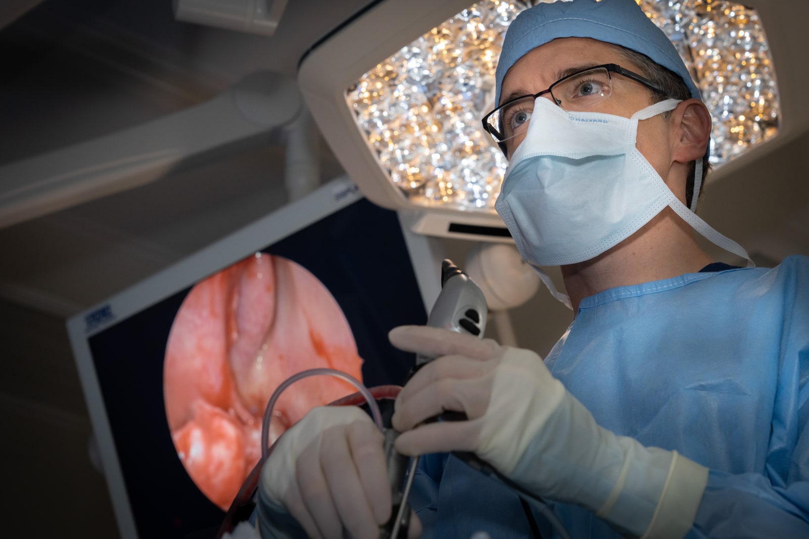
Nasal Endoscopy for Direct Visualization
A nasal endoscope allows physicians to examine the nasal cavity in real time. It reveals:
- Polyp size and position
- Drainage patterns
- Active inflammation
This tool guides both diagnosis and follow-up care.
Surgical Approaches in Complex Cases
Endoscopic Sinus Surgery
Modern sinus surgery is typically performed endoscopically. This means no external incisions are required. Small cameras and instruments are inserted through the nostrils.
In complex or revision cases, surgeons focus on:
- Restoring natural drainage pathways
- Removing polyps thoroughly
- Preserving healthy tissue
Improved visualization allows precise work near delicate skull base structures.
Image-Guided Navigation
In advanced cases, image-guided systems function like GPS for the sinuses. They connect surgical instruments to the patient’s CT scan in real time.
This technology improves safety when operating near:
- The eyes
- The brain
- Major blood vessels
Navigation systems are especially helpful in revision surgery.
Managing Chronic Inflammation Long Term
Surgery Is Only Part of Treatment
Removing polyps or opening blocked sinuses does not eliminate the underlying inflammatory condition. Long-term management is crucial.
Patients may require:
- Daily saline rinses
- Steroid nasal sprays
- Allergy treatment
- Asthma management
Surgery creates space for medications to work more effectively.
Minimally Invasive Office Procedures
For some patients, less invasive procedures may help manage chronic nasal obstruction without full sinus surgery. Treatments that target overactive nasal nerves can reduce congestion and drainage.
These newer approaches complement traditional sinus care in selected cases.
Recognizing Warning Signs of Skull Base Involvement
Patients should seek prompt evaluation if they experience:
- Clear, persistent nasal drainage on one side
- Vision changes
- Severe headaches
- Recurrent infections that do not respond to antibiotics
Early detection reduces the risk of serious complications.
Measuring Success After Complex Sinus Care
Improved Breathing and Smell
One key marker of successful treatment is restored airflow. Patients often report:
- Easier breathing
- Better sleep
- Reduced facial pressure
Smell may gradually return as inflammation decreases.
Fewer Infections and Medication Dependence
Effective treatment reduces the need for repeated antibiotics or oral steroids. This improves overall quality of life.
Ongoing Monitoring
Chronic sinus disease requires follow-up. Regular visits allow early detection of:
- Polyp regrowth
- Scar formation
- Recurrent inflammation
Long-term care supports lasting results.
Conclusion: Understanding the Bigger Picture of Sinus Disease
The nose, paranasal sinuses, and anterior skull base are closely connected. Because of this shared anatomy, disorders that begin with simple congestion can sometimes involve deeper structures.
Advanced imaging, precise surgical techniques, and long-term inflammatory control are essential in complex and revision cases. With tools like CT scans, nasal endoscopy, and image-guided surgery, specialists can safely manage challenging conditions.
Most importantly, patients benefit from early evaluation and comprehensive care. By understanding how these regions interact, individuals can seek timely treatment and protect both sinus function and overall health.